










Preoperative Planning with 3D Printed Anatomical Models

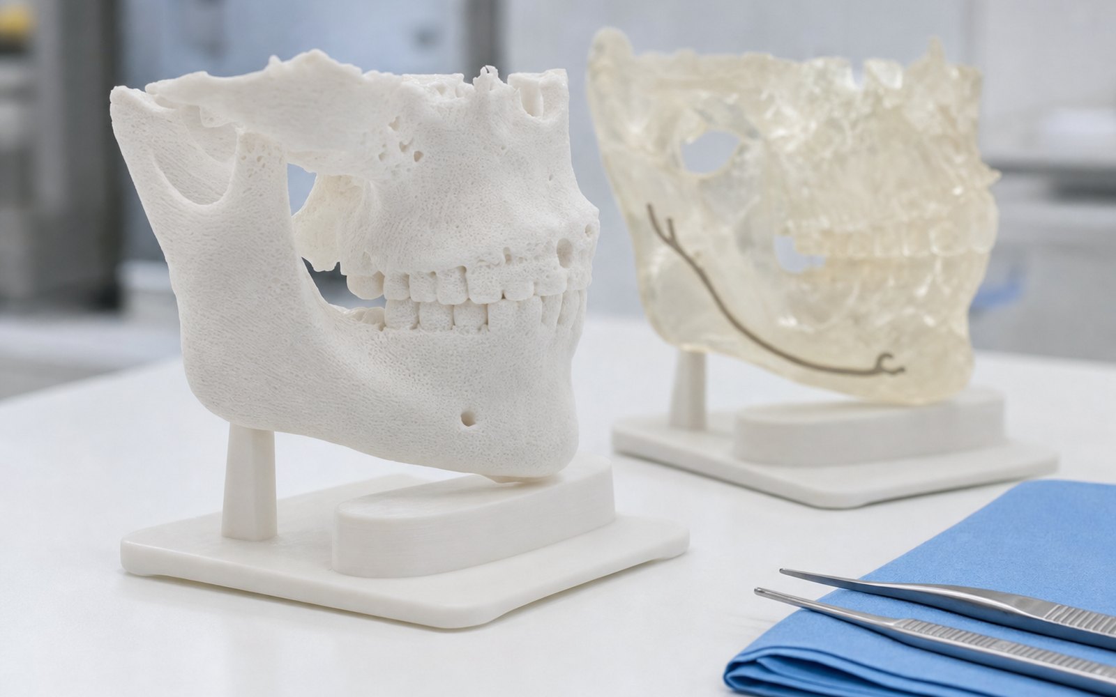
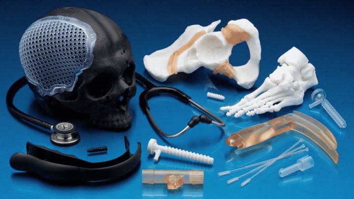
3D printed anatomical models turn a CT or MRI scan into an object a surgeon can hold, cut, drill and plan on before the patient is in the theatre. For a complex fracture, a deformity correction, a tumour resection or a revision hip, that rehearsal is the difference between a plan and a guess. The models are printed in polymer, cost a few thousand rupees, and are ready in days. This is how the process works, what the evidence says, and what to send us.
From scan to model
The workflow has four steps, and the first is the one that decides the quality of the model.
- Imaging. A CT for bone, an MRI for soft tissue, ideally with a slice thickness of 1 mm or less. A scan taken at 3 mm slices gives a model with steps in it; if the case matters, ask radiology for a thin-slice protocol.
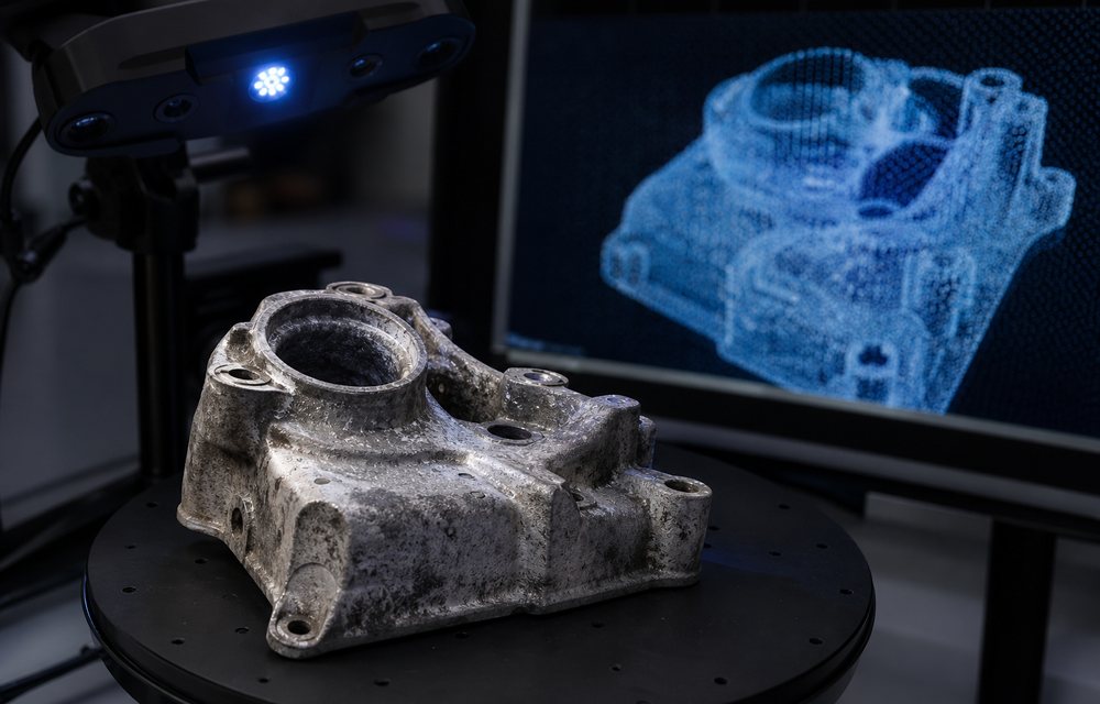
- Segmentation. The anatomy of interest is isolated from the scan, slice by slice, and turned into a surface mesh. This is done in software such as the free, open-source 3D Slicer, by the hospital’s radiology or biomedical team, or by us from the DICOM files.
- Preparation. The mesh is cleaned, scaled 1:1, and where needed split or hollowed for printing. A pelvis is printed whole; a skull may be printed in halves to show the interior.
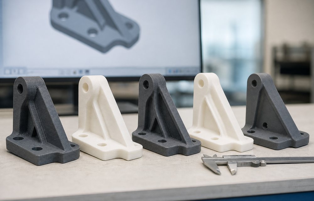
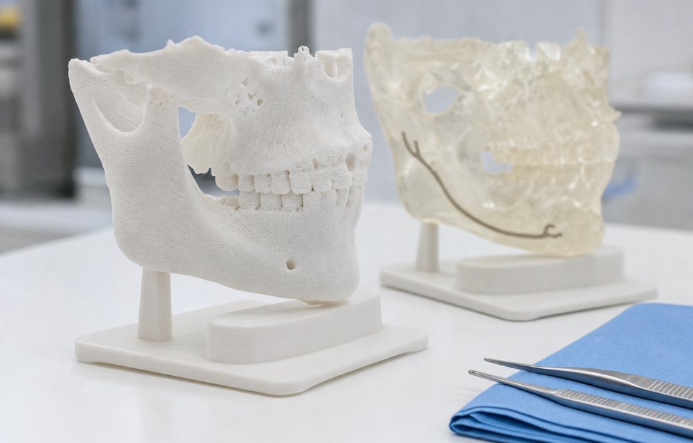
- Printing. FDM in PLA for most bone models: rigid, accurate, cheap, and it can be drilled and cut with the same tools used in theatre. SLA resin where fine detail or translucency is needed, for example a vascular tree inside a translucent kidney, or a middle ear.
What surgeons use the models for
- Choosing the approach. Turning a model in the hand shows a fracture pattern or a tumour’s relationship to a vessel in a way that scrolling through slices does not.
- Rehearsing the procedure. Osteotomies are cut on the model, screws are placed, and the plan is tested before it is tried on the patient.
- Pre-contouring implants. A plate bent to the model in the office fits the patient in theatre, which shortens the operation and reduces the number of times a plate is bent and re-bent.
- Consent and explanation. A patient who has held a model of their own anatomy understands the operation. Consent conversations get shorter and clearer.
- Teaching. Residents rehearse on the model, and rare cases become teaching material.
What the evidence says
Systematic reviews of 3D printing in surgery report reduced operating time and reduced blood loss in complex cases planned on a model, along with better communication with patients, with the strongest evidence in orthopaedic trauma, maxillofacial and cardiac surgery; a widely cited review by Tack and colleagues (2016) summarises the field. The honest caveat is that the largest savings are in the most complex cases, and that a routine procedure gains little. A model is for the case the surgeon is worried about.
Cost and turnaround
A typical bone model prints for between ₹5,000 and ₹15,000 depending on size and process, with a pelvis at the upper end and a hand or a mandible at the lower. Turnaround from a segmented file is two to four days; from raw DICOM it adds a day for segmentation. Urgent cases are handled faster, and we say at the outset if a scan is too coarse to give a model worth trusting.
Set against the cost of theatre time, a model that saves twenty minutes has paid for itself; one that avoids a second procedure has paid for a year of them.
Beyond the model: guides and implants
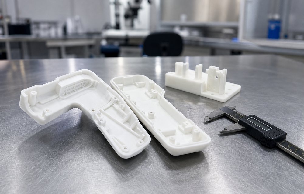
The model is the first step. From the same segmented anatomy, a surgical guide can be designed that sits on the patient’s bone and directs a drill or saw to the planned position; those are printed in a sterilisable biocompatible resin. Patient-specific implants are the step after that, and they are regulated devices with a development road of their own; our healthcare article sets out the four levels and the regulatory position of each.
Sending us a case
Send either the segmented STL or the anonymised DICOM series through the quote page, with the region of interest, the date of the procedure and whether the model will be cut or drilled. Patient data is handled under a written agreement and deleted after the job. The medical models page has examples and the materials we use.